Spondylolysis, Spondylolisthesis, Spondylitis & Spondylosis
These four conditions affect the lumbar spine and may be confusing. What is the difference between spondylolysis, spondylolisthesis, spondylitis, and spondylosis?
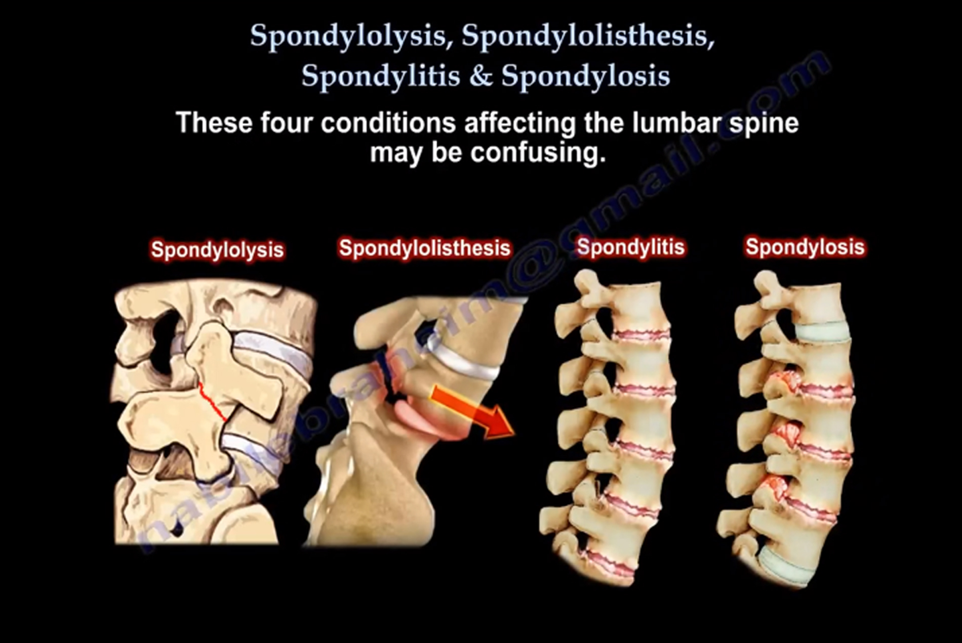
Spondylolysis (pars interarticularis Defect)
Used to describe the anatomic defect or break of the pars interarticularis of the vertebral arch. Spondylolysis usually occurs in the lower lumbar spine, especially the L5 vertebrae. It usually appears as a radiolucent gap on lateral X-ray. It occurs in about 5% of the population; it is not present at birth but develops over time and may run in families.
Spondylolysis is usually caused due to repetitive trauma, especially hyperextension. Repetitive trauma may cause stress fractures. The condition is usually asymptomatic; patient may have activity related low back pain or hamstring tightness. AP and lateral X-rays may show defect in about 80% of cases. It may show as a defect with or without sclerosis.
Oblique views may add another 15% more to the diagnosis. The "scotty dog" sign refers to the normal appearance of the lumbar spine, when seen on oblique radiographic projection. If spondylolysis is present, the pars interarticularis, or the neck of the dog, will have a defect or break (as if the dog has a collar around the neck).
CT scan is the best study, especially to check for healing. A SPECT scan is the best study when X-rays are negative and strongly suspect the condition. Lysis means dissolve or defect from a stress and it may be occult.
Spondylolisthesis (vertebral slippage)
Vertebra can start to shift out of place if the stress fracture weakens the bone and it is unable to maintain proper positioning. This forward slipping of the vertebrae may affect the nerves. It is not a disc slipping, but the slipping of the vertebral body. Another example is the Hangman's fracture (slippage of C2-C3). 15% of patients with pars defects progress to forward slippage. L5-S1 slippage occurs in 90% of pediatric cases. L4-L5 slippage is usually degenerative and usually occurs in female adults.
Large slip - continues to slip. Dysplastic slip - continues to progress.
- Congenital
- Isthmic - Most common: usually occurs due to pars defect (L5 - common)
- Degenerative - Occurs as a result of facet arthritis; Females older than 50 yrs.; African Americans; Usually affects the L4-L5 level; Typically the slip is not bad and rarely exceeds 30%; Commonly associated with instability and lumbar stenosis. May need flexion extension X-ray views to check for instability.
- Traumatic
- Pathologic
- Post-Surgical
- Type I (less than 35%)
- Type II (25-50%)
- Type III (50-75%)
- Type IV (75-100%)
- Type V (spondyloptosis)
Surgical Consideration in Treatment
L1-L4 - Repair defect if conservative treatment fails.
L5-S1 - In situ fusion in lower grades. - In high grade isthmic slippage, fusion from L4-S1. - Reduction of the slip may cause L5 nerve root injury.
Spondylitis (vertebral inflammation)
May affect the lower spine or the cervical spine.
- Pott's Disease (TB of the spine)
- Ankylosing Spondylitis - An autoimmune disease involving the spine and sacroiliac joints, and is also a form of spondylarthritis. The spine goes from stage of inflammation to stage of fusion. "Bamboo spine" is a radiographic feature that occurs as a result of vertebral body fusion by marginal syndesmophyte. HLB-27 is positive in the majority of cases.
Fractures in Ankylosing Spondylitis: Fractures may be occult. Get CT scan or MRI for the diagnosis. Be careful about neurological injury.
Spondylosis (vertebral arthritis)
The degenerative osteoarthritis of joints between the vertebrae and/or neural foramina. The space between the two adjacent vertebrae narrow which leads to compression of the nerve roots. Radiculopathy (compression of the nerve roots) will lead to pain, sensory, and motor change.
In the cervical spine, compression of the spinal cord from arthritis can lead to myelopathy.
Click the link below for my YouTube video on these four conditions:
https://www.youtube.com/watch?v=sHdAGSa1Opc
For more videos check out my YouTube channel: