
As a scientific conference approaches, I always think back to my first science fair: I stood next to my homemade presentation of graphs and tables glued to a poster board positioned next to hundreds of others made by fellow elementary school students. An overweight Paisan from rural New Jersey, I was more looking forward to my post-presentation reward of cannoli from my favorite bakery in South Philly than I was to standing for hours on end answering questions from the judges.
Conferences these days are pretty similar to the science fairs from decades ago, just on a grander scale with better technology. For example, every year the Organization for Human Brain Mapping (OHBM) brings together thousands of scientists who work with brain imaging data from around the world to share hot off the press findings that they just published or are preparing to publish. This year, 4,391 presentations will be on hand in Geneva for our annual conference. While there, thousands of us will stand next to posters as I did so many years ago, and a few lucky ones will be given the opportunity to present their work in front of a larger audience.
Most of these presentations are 12-15 minute bite-sized versions of a neuroimaging research project, which could easily be expanded to a 45-60 minute lecture. At the OHBM conferences, we have longer lectures too. Our Program Committee chooses outstanding speakers to summarize their own work in the context of the diverse field of brain mapping. This diversity is reflected in the wide range of topics at the conference spanning spatial scales (from single neurons to entire systems), behaviors (our senses, decision making, aging, consciousness, development, etc.), populations (healthy typical humans, populations with different clinical or psychiatric disorders, etc.) and of course, applications (computational models, artificial intelligence, etc).
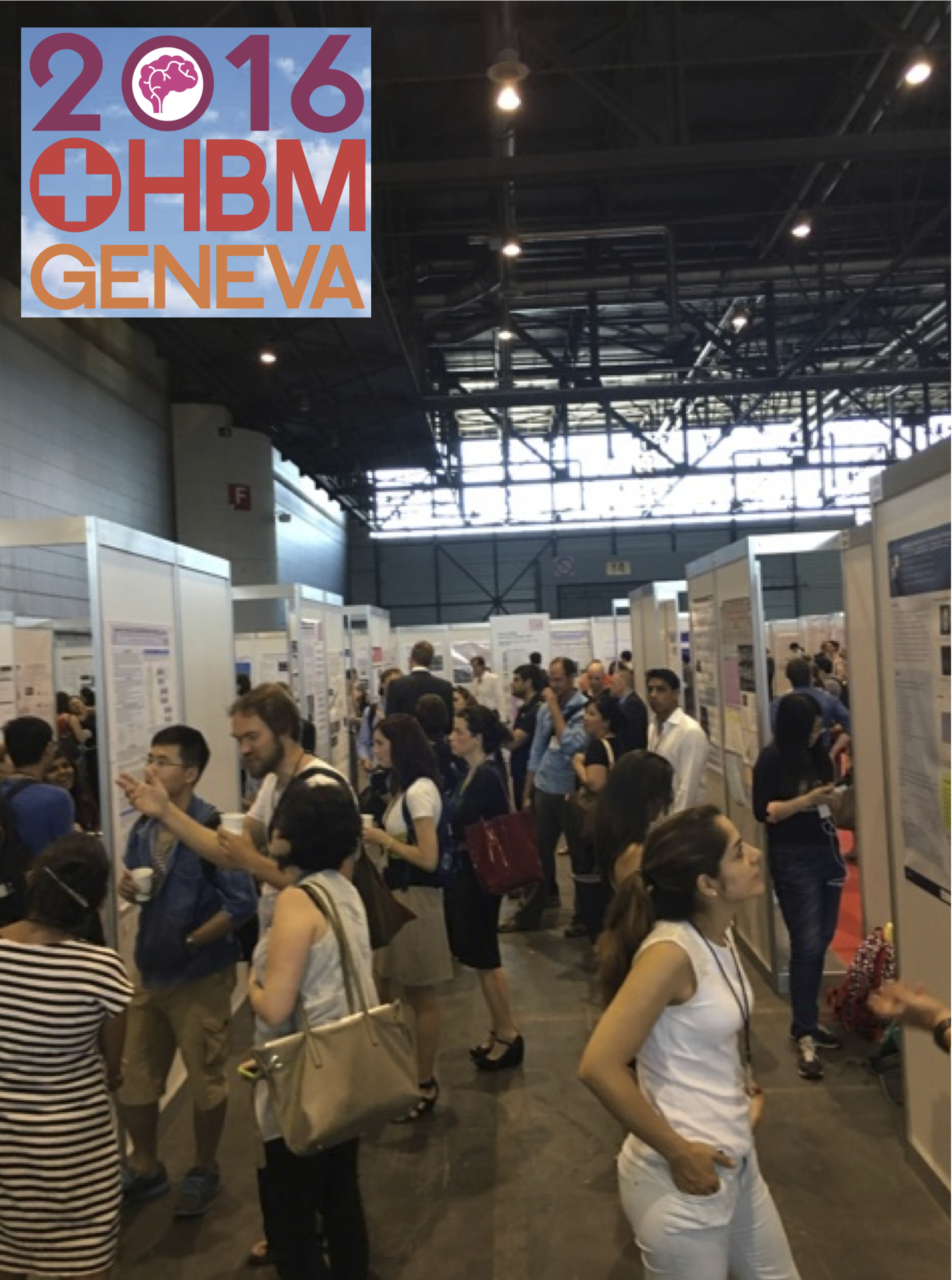
Brain mappers looking at posters this afternoon during this year's Organization for Human Brain Mapping meeting in Geneva.
For me, I'm a huge fan of the fusiform gyrus - so much so that at 33, I've already spent a third of my life staring and measuring this spindle-shaped brain structure. It's probably unsurprising then that I'm looking forward to learning about new ways to measure and quantify the fusiform in the living human brain at this year's meeting. The fusiform is interesting because it's involved in human-specific aspects of cognition such as reading and functional and structural abnormalities of the fusiform have been linked to disorders like autism.
The fusiform aside, conferences aren't all about being bombarded with data. Instead, they are also about re-connecting with people from the field who you know and setting foundations for new collaborations over a coffee or a pint or even a hike. Discussions of future collaborations also happen between organizations. For example, as Geneva is the home base for the World Health Organization (WHO), both organizations took the opportunity to set up a jointly held symposium between OHBM and WHO. Representatives from the major international brain initiatives will speak to raise awareness about current research projects that impact the staggering burden of brain disorders, and what we all might be able to do to reduce these burdens in the future. After all, understanding how the brain works is a team effort - no one can do it alone.
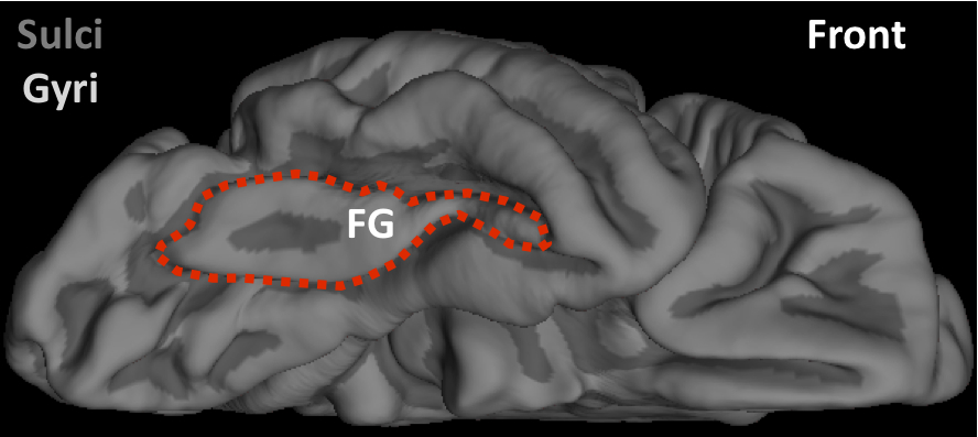
This is a cortical surface reconstruction of a right hemisphere averaged across 39 adults. This image is from FreeSurfer, which is an open source software package that you can download here. You are looking at the underside of the brain, which is rotated so the back is in the left of the image and the front in the right of the image. The fusiform gyrus (FG) is outlined in red. Fusiform is latin for spindle-shaped. Emil Huschke labeled this structure in 1854 because it is wider in the middle and tapers off at the ends like a spindle.
We also can't expect every single researcher at the meeting to blog about their presentations. And that's where Kat, Tzipi, Brendan, Emma, and I come in. As members of the OHBM Communications Committee, our boots (or for me, flip flops) are on the ground as we listen to the poster presentations. Our butts are in the chairs as we listen to the lectures. All the while, our fingers - the same ones that write the code to analyze our own data and that write our own empirical papers - will be typing away to come back and report to you what happened at the OHBM conference and at the meetings between OHBM and WHO.
And it's not just the five of us. There's a whole team of us. For example, we have already begun to post interviews with a subset of this year's speakers on our blog with more to come. Additionally, content from this year's meeting will soon be uploaded to our main site, which already contains lectures and courses from past meetings.
We are energized by the science and the thrill of discovery, and we love sharing these discoveries with all of you. And that's the main reason why we write and blog - to provide you with foundational information and up-to-date findings about the brain at the click of a button. We hope our posts give you insight into how your brain works and how to become informed consumers of neuroimaging news.